A Canon fundus camera is a specialized type of camera used to photograph the interior of the eye. It takes high-resolution photographs that allow for detailed examinations and diagnoses. The images are often compared to other medical imaging tests, such as magnetic resonance imaging (MRI) or computed tomography (CT).
Fundus cameras can be handheld or mounted on a tripod and use specific lenses to focus on different parts of the eye, such as the macula or optic nerve head. They can also capture multiple images at once with up to 70 degrees field of view. Additionally, these cameras are able to record digital video for further analysis, which makes them especially useful in diagnosing retinal diseases like diabetic retinopathy or macular degeneration.
The Canon Fundus Camera is a must-have tool for any ophthalmologist. It offers unparalleled accuracy, allowing doctors to get the most accurate readings of their patients’ eyes. This camera also allows practitioners to take high-resolution photographs and videos of the retina, which can be used for diagnostics and analysis.
With its advanced design and features, this camera provides professionals with a reliable diagnostic tool for eye care.
Table of Contents
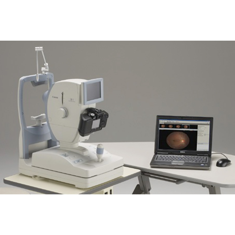
MED1400 EC Canon CR 2 Plus AF and imageSPECTRUM Capture Plus Tutorial
What is a Fundus Camera Used For?
A fundus camera is an ophthalmic imaging device that allows for the examination of a patient’s retina, optic nerve, and other structures at the back of their eye. It works by shining a light into the eye while taking pictures or video footage allowing ophthalmologists to assess and diagnose various vision problems such as glaucoma, macular degeneration, retinal detachment and diabetic retinopathy. Fundus cameras can also be used to monitor disease progression and treatment response over time.
Additionally, it is often used for educational purposes in medical schools providing students with an opportunity to observe different parts of the eye up close.
What is the Best Fundus Camera?
When choosing the best fundus camera, it is important to consider your specific needs and budget. The NIDEK AFC-210 is one of the most popular choices among ophthalmologists, thanks to its ultra-wide field of view (up to 200 degrees), high resolution imaging capabilities, and advanced software features that make capturing images easier than ever before. This model also includes a built-in LED light source for enhanced image quality and compatibility with other digital devices such as smartphones or tablets.
Additionally, the NIDEK AFC-210 offers an optional autofocus system that can be used for faster image acquisition in busy clinical settings. With all these features at an affordable price point, this fundus camera is definitely worth considering when searching for the best option on the market today.
Is Retinal Imaging Same As Fundus Photography?
Retinal imaging and fundus photography are both methods of capturing images of the back of the eye, but they differ in terms of the type and quality of images produced. Retinal imaging is a more advanced technology that uses digital cameras to capture high-resolution cross-sectional scans of the retina. These scans allow for detailed analysis by medical professionals, which can be used to diagnose various retinal diseases or disorders.
Fundus photography, on the other hand, is a more basic technique that captures still photographs using specialized lenses and lighting equipment. While these photographs may not provide as much detail as a scan produced with retinal imaging technology, they can still be useful in monitoring changes over time or diagnosing certain conditions such as diabetic retinopathy or macular degeneration.
What are the Different Types of Fundus Cameras?
A fundus camera is an imaging device used to take pictures of the interior surface of the eye, including the retina and choroid layer. There are several types of fundus cameras available on the market today. The two main categories are indirect ophthalmoscopy (using a handheld lens) and direct digital fundus photography (using a specialized digital camera).
Indirect ophthalmoscopy requires that an experienced practitioner manipulate a hand-held lens in order to capture images through dilated pupils, whereas direct digital fundus photography utilizes advanced optics and sophisticated software to quickly capture high quality photographs with minimal effort. Some models also offer features such as autofocus, image stabilization, color vision testing capabilities, contrast enhancement options, and other unique features. Depending on your needs and budget there is sure to be a suitable model for you!
Credit: www.digitaleyecenter.com
Canon Fundus Camera Price
The Canon Fundus Camera is an advanced medical imaging device used to take photographs of the back of a patient’s eye. It typically retails for around $20,000 USD, and packages may include additional equipment such as lenses, accessories and software. When purchasing this specialized camera system, it is important to make sure that you are getting all the necessary components to ensure accurate results and optimal performance.
Canon Cr2 Retinal Camera Manual
The Canon CR2 Retinal Camera Manual is an essential resource for anyone looking to operate the camera correctly and get the best quality images. This manual provides detailed instructions on how to use the CR2 Retinal Camera, as well as troubleshooting advice and safety tips. It also includes information about the various lenses available and their optimal settings for capturing stunning retinal photos.
Taking advantage of this manual will ensure that you can make the most of your Canon CR2 Retinal Camera!
Canon Cx-1
The Canon CX-1 is a 35mm film SLR camera originally released in 1983. It features an advanced program autoexposure system, which makes it easy to operate and ensures accurate exposure settings for a variety of situations. The CX-1 also has various manual controls, allowing photographers to customize their shooting experience.
Its viewfinder provides clear viewing with the help of its diopter adjustment lever and split image focusing screen. As one of the first cameras from Canon to feature programmed autoexposure and built-in motor drive capabilities, the CX-1 continues to be a popular choice for those looking for an affordable yet powerful film SLR camera.
Canon Cr2 Retinal Camera Price
The Canon Cr2 Retinal Camera is a top-of-the-line piece of medical equipment used to capture images of the retina. It offers high resolution, wide field of view, and advanced imaging capabilities. The cost for this camera varies depending on what version you purchase, but generally it ranges from $30,000-$60,000 USD.
This price tag reflects the level of quality and performance that comes with using this specialized device in your practice.
Canon Cx-1 Brochure
The Canon Cx-1 Brochure is a comprehensive guide to the features and benefits of the Canon Cx-1 camera. It provides detailed information on all aspects of its operation, from lens selection and image stabilization to built-in Wi-Fi connectivity and more. The brochure also includes a list of compatible accessories for further customization options, making it an ideal companion for anyone looking to get the most out of their photography experience with this powerful camera.
Canon Cr-2 Retinal Camera Software
The Canon CR-2 Retinal Camera Software is a specialized imaging software designed to help medical professionals capture, analyze and document retinal images. This tool helps with accurate image documentation for diagnosis and treatment of eye diseases such as macular degeneration, diabetic retinopathy, glaucoma and more. With features like automatic focus tracking and auto flash intensity control, the Canon CR-2 Retinal Camera Software makes capturing these delicate structures easy and efficient.
Canon Cr-2 Af Retinal Camera
The Canon CR-2 AF Retinal Camera is a specialized digital imaging device designed for clinical use in ophthalmology. It’s equipped with advanced features, like an auto focus system that quickly and accurately captures high-resolution retinal images. The camera also has an ergonomic design which makes it comfortable to use during long examination sessions.
This powerful piece of equipment provides quick and accurate diagnosis of various eye conditions and diseases, making it an invaluable tool in the fight against blindness.
Canon Cx-1 Manual
The Canon CX-1 Manual is an invaluable resource for anyone looking to get the most out of their Canon CX-1 camera. It provides comprehensive information on all aspects of the camera, from basic operation to more advanced techniques and settings. It also includes troubleshooting tips and detailed technical specifications so users can better understand how the camera works and make sure they are getting optimal results.
Conclusion
Overall, the Canon Fundus Camera is a great tool for investigating and diagnosing eye diseases. It has excellent image quality, easy-to-use technology, and advanced features that make it an ideal solution for ophthalmologists. In addition to being cost effective and versatile enough to use in most clinical settings, this camera offers superior performance when compared to other models in its class.
With this camera’s powerful capabilities, practitioners can confidently diagnose patients with greater accuracy than ever before.